42 eye diagram with labels and functions
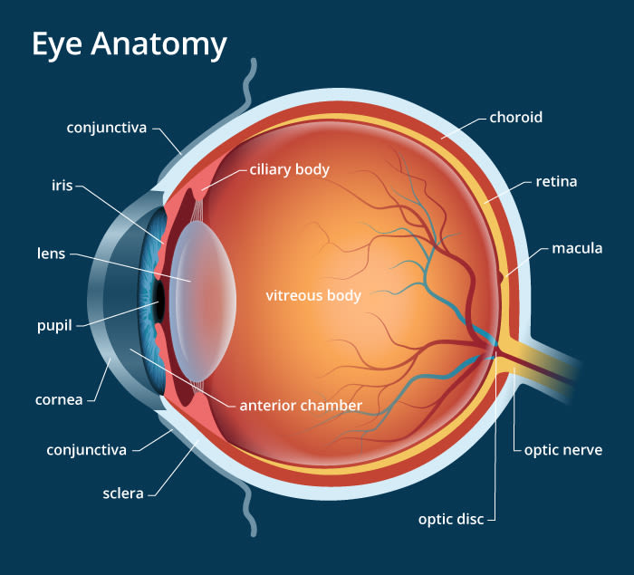
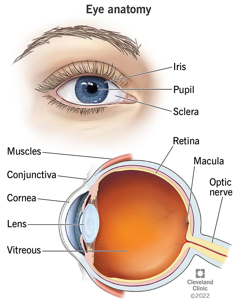
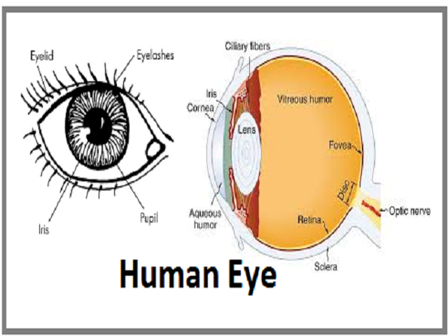
Labelled Diagram of Human Eye, Explanation and Function - VEDANTU The basic functions of Rods and Cones are conscious light perception, color differentiation and depth perception. The human eye is capable of distinguishing between about 10 million colors, and it can also detect a single photo. The human eye is a part of the sensory nervous system. Labeled Diagram of Human Eye Human Eye Diagram, How The Eye Work -15 Amazing Facts of Eye First, light rays enter the eye through the cornea, the clear front "window" of the eye. The dome shaped cornea bends light to help the eye focus. From the cornea, the light passes through an opening called the pupil. The amount of light passing through is controlled by the iris, or the colored part of your eye.
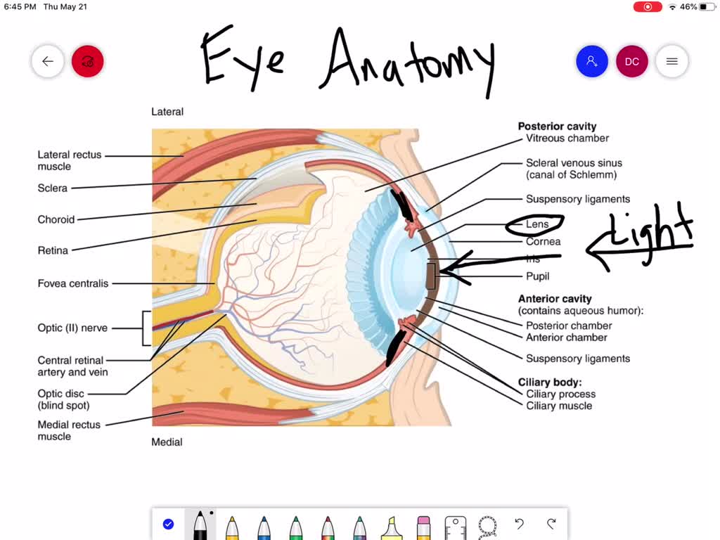
Eye anatomy: Muscles, arteries, nerves and lacrimal gland - Kenhub Six extraocular muscles move the eye: superior rectus, inferior rectus, medial rectus, lateral rectus, superior oblique and inferior oblique muscles; and one other, levator palpebrae superioris, opens the eyelid. Don't understand how all these muscles work? You can find out everything about them in the following learning materials.
Eye diagram with labels and functions
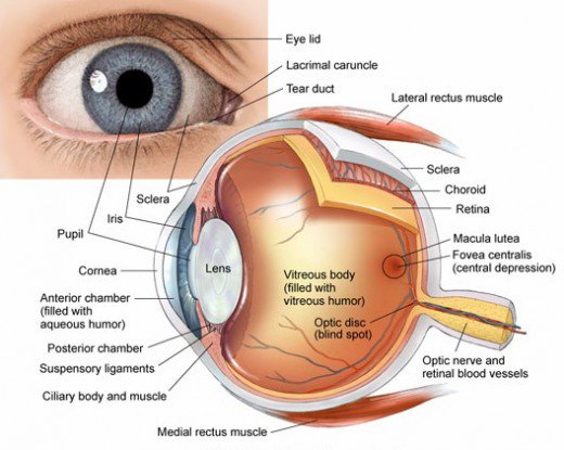
Structure of Human Eye (With Diagram) | Human Body - Biology Discussion The function of the eyebrows is to protect the anterior aspect of the eyeball from sweat, dust and other foreign bodies. 2. The Eyelids (Palpebrae) and Eyelashes: The eyelids are two movable folds situated above and below front of the eye. On their free edges, there are outgrowths of hairs— the eyelashes. Diagram of the Eye - Lions Eye Institute To understand the eye and its functions, it's important to understand how the eye works, see below diagrams for both the external eye and the internal eye. The External Eye Instructions Click the parts of the eye to see a description for each. Hover the diagram to zoom. The Internal Eye Instructions The Eye Diagram: What is it and why is it used? The eye diagram is used primarily to look at digital signals for the purpose of recognizing the effects of distortion and finding its source. To demonstrate using a Tektronix MDO3104 oscilloscope, we connect the AFG output on the back panel to an analog input channel on the front panel and press AFG so a sine wave displays. Then we press Acquire.
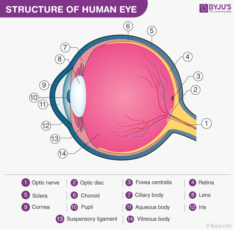
Eye diagram with labels and functions. Structure and Functions of Human Eye with labelled Diagram - BYJUS Structure and Functions of Human Eye with labelled Diagram Biology Biology Article Structure Of Eye Structure of the Eye The eye is one of the sensory organs of the body. In this article, we shall explore the anatomy of the eye The structure of the eye is an important topic to understand as it one of the important sensory organs in the human body. Anatomy of the eye: Quizzes and diagrams | Kenhub Take a look at the diagram of the eyeball above. Here you can see all of the main structures in this area. Spend some time reviewing the name and location of each one, then try to label the eye yourself - without peeking! - using the eye diagram (blank) below. Unlabeled diagram of the eye. Click below to download our free unlabeled diagram of ... Labelling the eye — Science Learning Hub In this interactive, you can label parts of the human eye. Use your mouse or finger to hover over a box to highlight the part to be named. Drag and drop the text labels onto the boxes next to the eye diagram If you want to redo an answer, click on the box and the answer will go back to the top so you can move it to another box. MCAT Eye Anatomy: Eye Structure & Function - Magoosh MCAT Blog MCAT Eye Anatomy: Diagram of the Human Eye Light refracts (bends) as it passes sequentially through the cornea, aqueous humor, lens, and vitreous humor. Errors in refraction cause visual defects which can be corrected by contacts or glasses. Myopia and hyperopia are two types of refractive error.
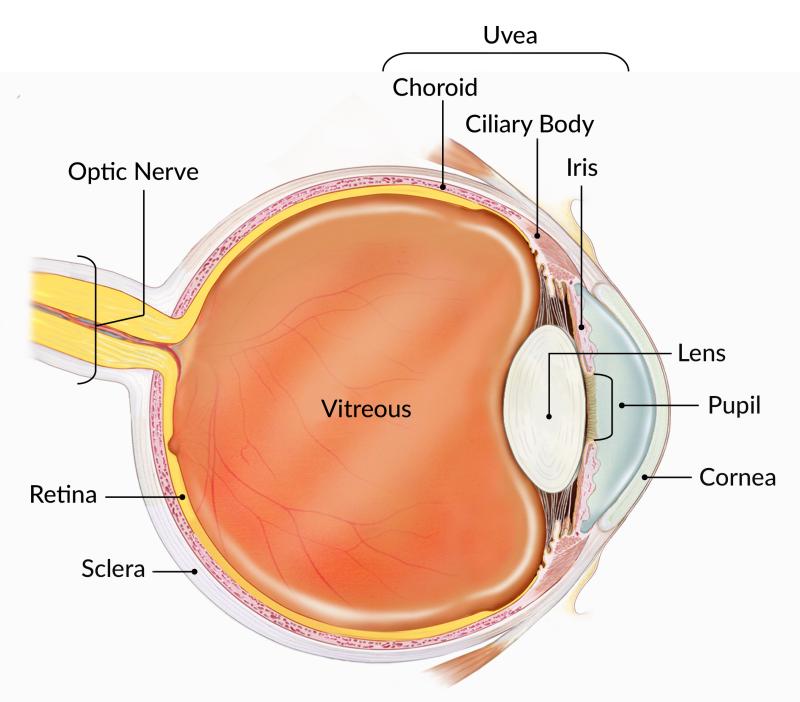
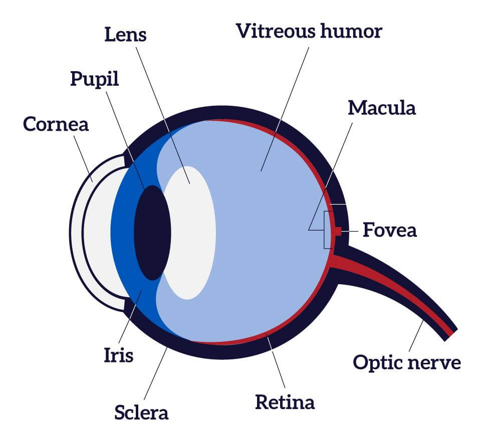
Eye Anatomy: A Closer Look At the Parts of the Eye - All About Vision For more details about specific structures of the eye and how they function, visit these pages: Conjunctiva Of The Eye. Sclera: The White Of The Eye. Cornea Of The Eye. The Uvea Of The Eye. Pupil: Aperture Of The Eye. The Retina: Where Vision Begins. Macula Lutea Of The Eye. Choroid Of The Eye. Lens Of The Eye. Ciliary Body. Eye Muscles ... Structure And Function Of The Eye - Vision - MCAT Content - Jack Westin The iris, which is conspicuous as the colored part of the eye, is a circular muscular ring lying between the lens and cornea that regulates the amount of light entering the eye. In conditions of high ambient light, the iris contracts, reducing the size of the pupil at its center. In conditions of low light, the iris relaxes and the pupil enlarges. Eye anatomy and function - AboutKidsHealth The pupil, or black dot at the centre of the eye, is an opening through which light can enter the eye. The iris, or coloured part of the eye, surrounds the pupil. It controls how much light enters the eye by changing the size of the pupil. The cornea, a clear window at the front of the eye, covers the iris and the pupil. Eye Parts Labeling and Functions Flashcards | Quizlet Start studying Eye Parts Labeling and Functions. Learn vocabulary, terms, and more with flashcards, games, and other study tools.
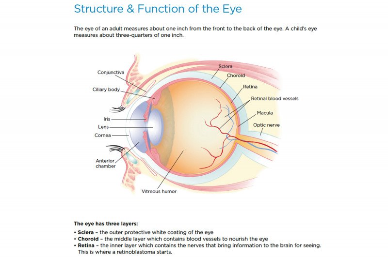
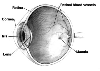
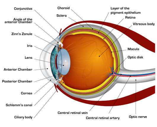
PDF Parts of the Eye - National Institutes of Health To understand eye problems, it helps to know the different parts that make up the eye and the functions of these parts. Here are descriptions of some of the main parts of the eye: ... Handout illustrating parts of the eye Keywords: parts of the eye, eye diagram, vitreous gel, iris, cornea, pupil, lens, optic nerve, macula, retina ... Eye Anatomy | Definition, Structure & Functions - iBiologia Diagram of Human Eye with Labelling. Eye Anatomy Complete Physiology of Eye is described below in the given paragraph: The eye is rather like a living Camera. Each eye is a liquid-filled ball 2.5 cm in diameter. At the front of the eye is a clear, round window called the cornea. Behind the cornea is a "lens. Eye Diagram With Labels and detailed description - BYJUS A brief description of the eye along with a well-labelled diagram is given below for reference. Well-Labelled Diagram of Eye The anterior chamber of the eye is the space between the cornea and the iris and is filled with a lubricating fluid, aqueous humour. The vascular layer of the eye, known as the choroid contains the connective tissue. Parts Of The Eye Labeled Diagram Model And Their Function Parts of the eye-labeled diagram model are divided into three groups: the external outer layer, the middle layer, and the inner back layer. The outer layer is responsible for protecting the eye from environmental toxins and debris. The middle layer includes cells that allow light to enter and travel through the back layer to the retina.
Eye Anatomy: A Closer Look At the Parts of the Eye
Cow's Eye Dissection - Eye diagram - Exploratorium The pupil is the dark circle in the center of your iris. It's a hole that lets light into the inner eye. Your pupil is round. A cow's pupil is oval. A tough, clear covering over the iris and the pupil that helps protect the eye. Light bends as it passes through the cornea. This is the first step in making an image on the retina.
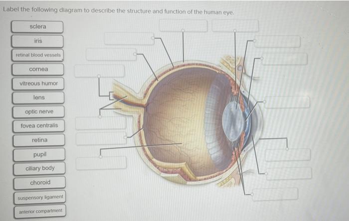
Solved Label the following diagram to describe the structure ...
Eye Anatomy: 16 Parts of the Eye & Their Functions - Vision Center The following are parts of the human eyes and their functions: 1. Conjunctiva The conjunctiva is the membrane covering the sclera (white portion of your eye). The conjunctiva also covers the interior of your eyelids. Conjunctivitis, often known as pink eye, occurs when this thin membrane becomes inflamed or swollen.

Structure And Functions of the different part of the Human ...
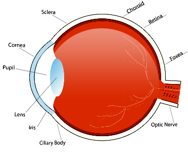
eye labeling Diagram | Quizlet sclera. Tough white out covering of the eyeball. choroid. Middle layer of the eye (between the retina and the sclera) that contains the blood vessels that nourish the eye and cornea. iris. colored layer that dilates and constricts to allow in more or less light. ciliary body. structure on each side of the lens that connects the choroid and iris.
Human Eye: Anatomy, parts and structure - Online Biology Notes
PDF Eye Anatomy Handout - National Institutes of Health of light entering the eye. Lens: The lens is a clear part of the eye behind the iris that helps to focus light, or an image, on the retina. Macula: The macula is the small, sensitive area of the retina that gives central vision. It is located in the center of the retina. Optic nerve: The optic nerve is the largest sensory nerve of the eye.
Label the following diagram of the eye and briefly state t ...
Eye Diagram Printable: Free Worksheet for Kids Eye Diagram Printable. 5.0 based on 81 votes. Ask any toddler where their eyes are, and someone is bound to get their eyes poked! Kids learn basic body parts early on, but older kids need to learn the intricacies of anatomy. Work with your child using this parts of the eye worksheet to help him or her learn more about their sense of sight!
Eyes: How They Work, Anatomy & Common Conditions
Label Parts of the Human Eye - University of Dayton Parts of the Eye. Select the correct label for each part of the eye. The image is taken from above the left eye. Click on the Score button to see how you did. Incorrect answers will be marked in red. ...

IGCSE Biology 2017: 2.91: Describe the Structure and Function ...
Labelling the eye — Science Learning Hub Activity Labelling the eye The human eye contains structures that allow it to perceive light, movement and colour differences. In this activity, students use online or paper resources to identity and label the main parts of the human eye. Citizen science Teacher PLD Glossary Sign in Email Us See our newsletters here. News and Events About
describe the anatomy of the eye and the function of each part pages 310 14
Label the Eye - The Biology Corner Label the Eye. Shannan Muskopf December 30, 2019. This worksheet shows an image of the eye with structures numbered. Students practice labeling the eye or teachers can print this to use as an assessment. There are two versions on the google doc and pdf file, one where the word bank is included and another with no word bank for differentiation.

30 Amazing Facts About Human Eyes For Kids, With Diagrams ...
Labeled Eye Diagram | Science Trends What you want to interpret as a major part of the human eye is somewhat up to the individual, but in general there are seven parts of the human eye: the cornea, the pupil, the iris, the lens, the vitreous humor, the retina, and the sclera. Let's take a closer look at each of these components individually. The Cornea

How your eye works (parts of the eye)Look After Your Eyes
labelled diagram of human eye - Microsoft eye labelled diagram human label labels draw eyes well labeling Human Eye: Anatomy, Structure And Function robotics maximillian selorm doku Diagram Showing Parts Of Human Eye 455677 Vector Art At Vecteezy eye diagram human parts showing illustration vector safety vecteezy

Parts and Functions of the Human Eye Diagram | Quizlet
Eye Anatomy Diagram - EnchantedLearning.com Retina - light-sensitive tissue that lines the back of the eye. It contains millions of photoreceptors (rods and cones) that convert light rays into electrical impulses that are relayed to the brain via the optic nerve. Rods - cells the in the retina that sense brightness (they are photoreceptors). Night vision involves mostly rods (not cones).

File:Three Main Layers of the Eye.png - Wikimedia Commons
The Eye Diagram: What is it and why is it used? The eye diagram is used primarily to look at digital signals for the purpose of recognizing the effects of distortion and finding its source. To demonstrate using a Tektronix MDO3104 oscilloscope, we connect the AFG output on the back panel to an analog input channel on the front panel and press AFG so a sine wave displays. Then we press Acquire.
How the Eyes Work | National Eye Institute
Diagram of the Eye - Lions Eye Institute To understand the eye and its functions, it's important to understand how the eye works, see below diagrams for both the external eye and the internal eye. The External Eye Instructions Click the parts of the eye to see a description for each. Hover the diagram to zoom. The Internal Eye Instructions
Retinoblastoma: Anatomy of the Eye | Memorial Sloan Kettering ...
Structure of Human Eye (With Diagram) | Human Body - Biology Discussion The function of the eyebrows is to protect the anterior aspect of the eyeball from sweat, dust and other foreign bodies. 2. The Eyelids (Palpebrae) and Eyelashes: The eyelids are two movable folds situated above and below front of the eye. On their free edges, there are outgrowths of hairs— the eyelashes.

Structure And Function Of The Eye - Vision - MCAT Content

Labeled Eye Diagram | Human eye diagram, Eye anatomy, Diagram ...

Anatomy of The Eye 101 | Eyecheck
Eyes (Anatomy): Overview, Parts and Functions | Biology ...

Human Eye Anatomy - Parts of the Eye Explained | Eye anatomy ...

15.5 Vision – Anatomy & Physiology
How the Human Eye Works | Cornea Layers/Role | Light Rays

Structure and Functions of Human Eye with labelled Diagram

Simple eye diagrams | Easy eye diagram | Labeled eye diagram ...

Parts of the Human Eye Diagram | Quizlet
/GettyImages-695204442-b9320f82932c49bcac765167b95f4af6.jpg)
Structure and Function of the Human Eye
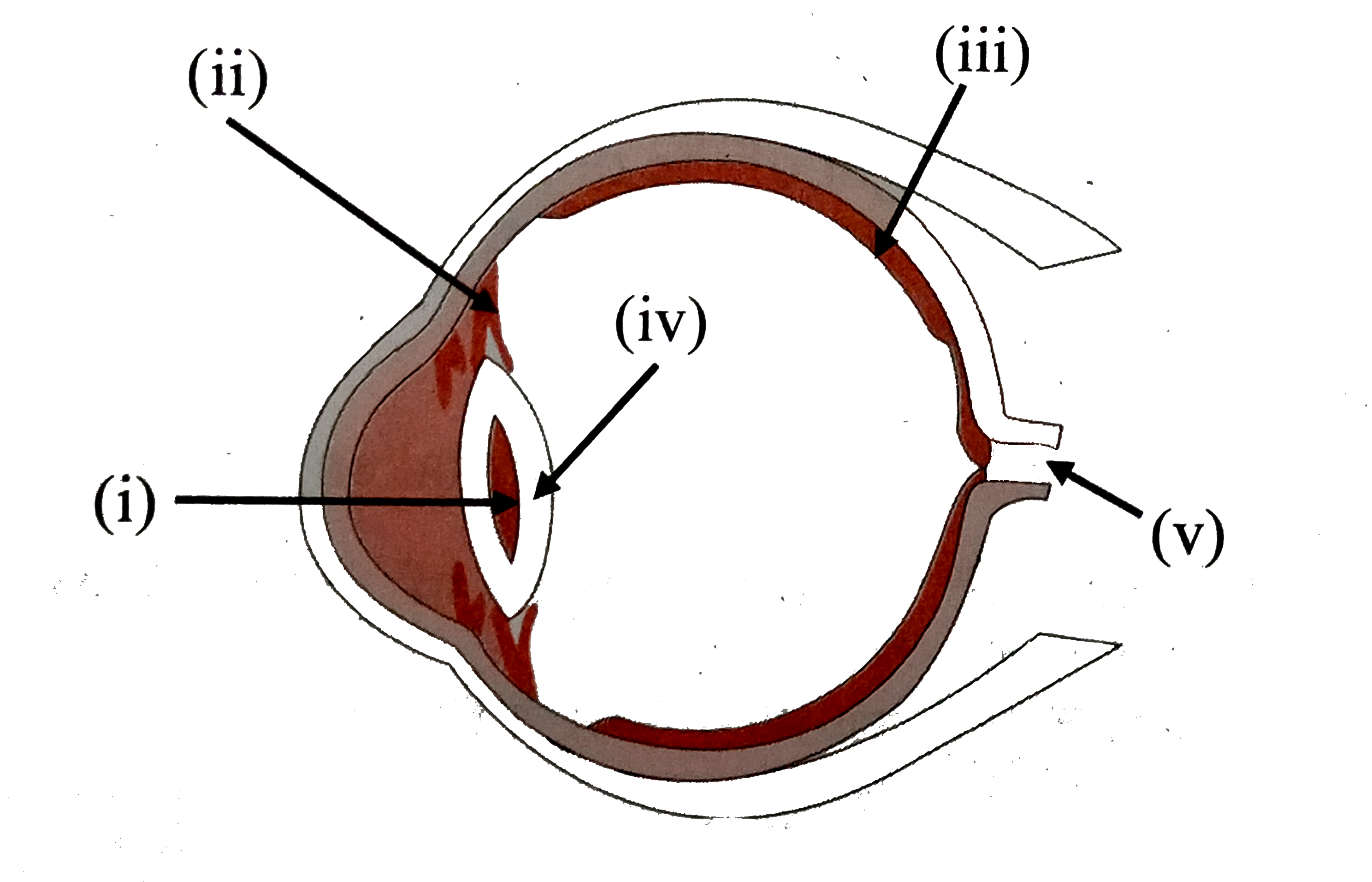
Label the parts of the following diagram of the human eye and ...

Label the Eye Diagram | Quizlet

Eye Anatomy and How the Eye Works

Eye diagram by Firkin | Human eye diagram, Diagram of the eye ...
Structure and Functions of Human Eye with labelled Diagram
The Human Eye: Anatomy, Structure, Working, Function and Defects

Human eye anatomy _Eye part and function

Human Eye Ball Anatomy & Physiology Diagram

Simple eye diagrams | Easy eye diagram | Labeled eye diagram ...
Human Eye Diagram, How The Eye Work -15 Amazing Facts of Eye

Anatomy of the Human Eye

Anatomy Of Eyes Teaching Resources | Teachers Pay Teachers

Anatomy of The Eye 101 | Eyecheck
Neuroscience for Kids - Fill In #3

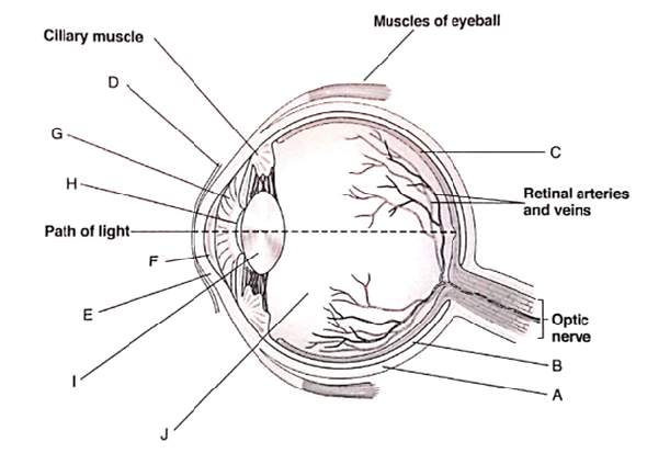
draw a neat labelled diagram of the human eye and mention the ...
Parts of the Eye & Their Function | Robertson Optical and ...

Pupil Function, Anatomy & Size | What Does the Pupil of the ...
Post a Comment for "42 eye diagram with labels and functions"