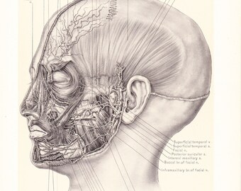
45 face diagram with labels
Learn the facial muscles with quizzes & labeled diagrams - Kenhub Simply fill in the blanks with the face muscles names you remember. DOWNLOAD PDF WORKSHEET (BLANK) DOWNLOAD PDF WORKSHEET (LABELED) How did you get on? If you still need a bit of practice, no worries. This was just a warm-up to get your brain thinking! The next stage is to test yourself using some interactive facial muscles quizzes. Parts of a microscope with functions and labeled diagram Figure: Diagram of parts of a microscope There are three structural parts of the microscope i.e. head, base, and arm. Head - This is also known as the body. It carries the optical parts in the upper part of the microscope. Base - It acts as microscopes support. It also carries microscopic illuminators.
Horse Skeleton Anatomy - Osteological Features of Bones from Equine ... Horse skeleton anatomy diagram Few special osteological features from the axial and appendicular skeleton of a horse - The skull of a horse is long and four-sided. You will find an extensive foramen lacerum in the horse skull. There is no cornual process in horse skull. The fusion between the two haves of the mandible is complete.
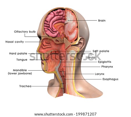
Face diagram with labels
ML | Face Recognition Using Eigenfaces (PCA Algorithm) We first convert these images into vectors of size N2 such that: Now we calculate the average of all these face vectors and subtract it from each vector average_face Now we take all face vectors so that we get a matrix of size of N2 * M. Now, we find Covariance matrix by multiplying A with AT. A has dimensions N2 * M, thus AT has dimensions M * N2. Face Recognition Homepage - Databases The MIT-CBCL face recognition database contains face images of 10 subjects. It provides two training sets: 1. High resolution pictures, including frontal, half-profile and profile view; 2. Synthetic images (324/subject) rendered from 3D head models of the 10 subjects. Anatomy of the face and neck (MRI) - e-Anatomy - IMAIOS The bones of the face and neck were labeled using different colors to facilitate comprehension. The bone structures are rather more difficult to view on a weighted MRI T2 than on a CT-Scan: for more details on the bones of the face, please refer to the e-Anatomy module "Face-CT-Scan". The teeth were numbered using the FDI World Dental ...
Face diagram with labels. Facial Nerve: Anatomy, Function, and Treatment - Verywell Health The facial nerve and its branches regulate a number of functions of the mouth and face. Most of its divisions stimulate muscles that allow eyelids to open and close, as well as facial movements. This nerve also mediates the production of tears and saliva and perception of taste in the tongue and receives some sensory input from the face as well ... Anatomical Position and Directional Terms: Definitions, Example Labeled ... We will first review the anatomical position, its definition, and look at example labeled diagrams. We will then walk through the different anatomical directional terms used to describe location and movement. We will provide you with labeled diagrams, example body parts, and tricks to learn the directional terms listed below! Medial vs Lateral Anatomical Line Drawings - Medscape Surface Anatomy - lateral views - male. go to drawing without labels. Surface Anatomy - lateral views - female. go to drawing without labels. Surface Anatomy - Child - anterior view & posterior ... Arteries Diagram Labeled Quizlet / Artery and Vein Model | Arteries and ... The veins of head and neck are labeled in blue . Start studying label the arteries. Veins of the head and neck: Start studying lab2, arteries and veins labeling. Learn how blood is pumped away from the heart at high pressure in arteries, and returns to the heart at low pressure in veins. Artery that supplies blood to the face, head, and neck.
WHMIS 2015 - Labels : OSH Answers Suppliers and employers must use and follow the WHMIS 2015 requirements for labels and safety data sheets (SDSs) for hazardous products sold, distributed, or imported into Canada. Please refer to the following other OSH Answers documents for more information: WHMIS 2015 - General. WHMIS 2015 - Pictograms. Human face: anatomy, structure and function | Kenhub This portion of the face is marked by four regions including the nasal, infraorbital, zygomatic and auricular regions. The nasal region is located in the central portion of the human face and, as its name suggests, it features the nose. The nose is the central pyramid-shaped structure, situated in the midline. Female Anatomy: Labeled Diagrams of the Reproductive System Female anatomy refers to the internal and external structures of the reproductive and urinary systems. Reproductive anatomy aids with sexual pleasure, getting pregnant, and breastfeeding a baby. The urinary system helps rid the body of toxins through urination (peeing). The Female Reproductive System. Some people are born with internal or ... Parts of A Check Labeled & Explained (with Diagrams) [2021] I'll also show you helpful diagrams with labels so that you can properly identify and understand each part. Table of Contents Parts of the check explained 1. Contact information 2. Date 3. Pay to the order of 4. Transaction amount 5. "Money box" 6. Memo 7. Signature line 8. Routing number 9. Account number 10. Check number Parts of a check FAQ
Entity Relationship Diagram (ERD) | ER Diagram Tutorial There are three basic elements in an ER Diagram: entity, attribute, relationship. There are more elements which are based on the main elements. They are weak entity, multi valued attribute, derived attribute, weak relationship, and recursive relationship. Cardinality and ordinality are two other notations used in ER diagrams to further define ... Muscles of Facial Expression | Anatomy | Geeky Medics These sections include the occipital (located posteriorly) and frontal (located anteriorly) bellies. The frontal belly is the major contributor to facial expression.¹ Origin The occipital belly originates from the occipital bone, as well as the mastoid process of the temporal bone. The frontal belly originates from the epicranial aponeurosis .¹ How to plot a ternary diagram in Excel - Chemostratigraphy.com Adding labels to the tick marks Use the Add Chart Element > Add Labels ( Chart Design tab) to add Data Labels to the A to B axis aligned to the right (Figure 17), then add Data Labels aligned left to the C to A axes. Figure 17: Adding Data Labels to the A-B and B-C axes. By default, Excel will use the Y Value as Data Label. Anatomy, Head and Neck, Face - StatPearls - NCBI Bookshelf The ears enclose the lateral borders of the central face. The central face region contains the nose, cheeks, and ears. Nose The nose is a midline structure that protrudes from the face. This protuberance is made from cartilage predominantly. But the base of the nose is made from the nasal bones.
Scan atlas of anatomy of the face - e-Anatomy - IMAIOS Anatomy of the face and neck (CT) - interactive atlas of human anatomy using cross-sectional imaging. This head and neck anatomy atlas is an educational tool for studying the normal anatomy of the face based on a contrast enhanced multidetector computed tomography imaging (axial and coronal planes). Interactive labeled images allow a ...
Microscope, Microscope Parts, Labeled Diagram, and Functions The description given below summarize the brief description of microscope parts used to visualize the microscopic specimens such as animal cells, plant cells, microbes, bacteria, viruses, microorganisms etc. The Microscopes parts divided into three different structural parts Head, Base, and Arms. Head/Body: It contain the optical parts in the ...

Highlighter is all about emphasizing the places where the light naturally hits your face. For a ...
Facial Bones Anatomy | List & Functions - Video & Lesson ... - Study.com This image of the skull shows the names of the 14 facial bones. The names of the 14 facial bones are: Inferior nasal concha x 2 Lacrimal bones x 2 Mandible Maxilla x 2 Nasal bones x 2 Palatine...
Circle Diagram: What It Is, Templates & Use Cases - Venngage 2. Select one of our professionally designed circular diagram templates or choose a blank canvas. 3. Start editing using our drag-and-drop editor or smart diagram editor. 4. Now's the fun part! Add or replace icons, illustrations or images to your heart's content. Beef up your circle diagram design with icons, illustrations and images.
How to label eyes detected using mediapipe - Stack Overflow To label the eye, first we need to find the left-right eye coordinate using. import cv2 import mediapipe as mp from mediapipe.python.solutions.drawing_utils import _normalized_to_pixel_coordinates dframe = cv2.imread ("person.png") image_input = cv2.cvtColor (dframe, cv2.COLOR_BGR2RGB) # load face detection model mp_face = mp.solutions.face ...
These Are the 12 Cranial Nerves and Their Functions - Healthline Your cranial nerves are pairs of nerves that connect your brain to different parts of your head, neck, and trunk. There are 12 of them, each named for its function or structure. Their functions ...
Ask a Dermatologist: How to Read Skincare Ingredient Labels Ingredients are listed from highest to lowest concentration, says Jacqueline Schaffer, an anti-aging expert, best-selling author, and founder of vegan skincare brand Schique. This means if a really great ingredient is listed at the bottom, you're not going to get much benefit from it. "Typically only the top 'active ingredients' are required to ...
Labeled imaging anatomy cases | Radiology Reference Article ... cerebral angiogram (DSA): lateral cerebral angiogram (DSA): frontal Head and neck CT head: bone window axial skull base CT head: bone window axial calvarium CT facial bones/orbits: axial CT facial bones/orbits: coronal CT facial bones/orbits: sagittal CT temporal bone: axial CT temporal bone: coronal CT temporal bone: sagittal
Anatomical Position: Body Planes and Sections - EZmed The sagittal plane is the green line labeled "S" on the image below, the coronal plane is the blue line labeled "C", and the transverse plane is the yellow line labeled "T". If we use the abbreviation SCT, you can also remember the first 2 planes are vertical (sagittal and coronal), and the last plane is horizontal (transverse).
Labeling Theory - Simply Psychology These labels are informal (Kavish, Mullins, and Soto, 2016). Labeling and Deviant Behavior Labeling can encourage deviant behavior in three ways: a deviant self-concept, a process of social exclusion, and increased involvement in deviant groups. Deviant Self-Concept Deviant self-concept originates from the theory of symbolic interactionism.
What are the 12 cranial nerves? Functions and diagram Scientists use Roman numerals from I to XII to label the cranial nerves in the brain. The 12 cranial nerves include the: olfactory nerve optic nerve oculomotor nerve trochlear nerve trigeminal...
Anatomy of the face and neck (MRI) - e-Anatomy - IMAIOS The bones of the face and neck were labeled using different colors to facilitate comprehension. The bone structures are rather more difficult to view on a weighted MRI T2 than on a CT-Scan: for more details on the bones of the face, please refer to the e-Anatomy module "Face-CT-Scan". The teeth were numbered using the FDI World Dental ...
Face Recognition Homepage - Databases The MIT-CBCL face recognition database contains face images of 10 subjects. It provides two training sets: 1. High resolution pictures, including frontal, half-profile and profile view; 2. Synthetic images (324/subject) rendered from 3D head models of the 10 subjects.
ML | Face Recognition Using Eigenfaces (PCA Algorithm) We first convert these images into vectors of size N2 such that: Now we calculate the average of all these face vectors and subtract it from each vector average_face Now we take all face vectors so that we get a matrix of size of N2 * M. Now, we find Covariance matrix by multiplying A with AT. A has dimensions N2 * M, thus AT has dimensions M * N2.





Post a Comment for "45 face diagram with labels"